
X-ray Imaging and its use in Life Science Research
Free Webinar | 19 March 2024Understanding physiological structures is at the core of many research questions in life science. Electron microscopy offers structural information at the ultra-resolution level, but you may wish to capture larger volumes from your samples. Or perhaps you would like to streamline your multimodal imaging workflows by quickly acquiring an overview 3D dataset.
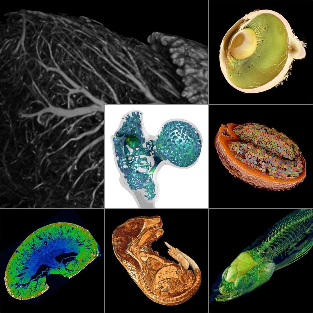
ZEISS X-ray microscopes provide high contrast, high resolution 3D imaging of your delicate biological samples including mineralized and soft tissues, organs and organoids, plant tissues and more. Study inside your specimen histologically, without destroying your sample with dissection, down to a cellular level.
Speakers

Rosy is a Solution Manager at ZEISS Microscopy and became fascinated by microscopy during her PhD when she used confocal microscopes extensively to explore preimplantation embryo development. In 2008 Rosy joined ZEISS UK as an imaging specialist focusing on the laser-based 3D instruments including confocal, super resolution and lightsheet microscopy. Rosy took responsibility for the X-ray microscopy portfolio in the UK in 2015 and was immediately struck by the vast range of specimens that can be explored, particularly in biological research. Having progressed through a number of global roles, Rosy now works in the life science division as a solution manager where she supports the global ZEISS team in developing applications, sharing expertise and defining future requirements for the ZEISS portfolio of X-ray instruments.

Mohsen Samadi Khoshkhoo is a business development manager at ZEISS for X-ray microscopy systems in the region Europe, the middle east and Africa (EMEA). With his BSc and MSc in metallurgy he worked for a year in a steel company followed by a year in a materials and energy research center in Iran. In 2009 he joined to Leibniz Institute for Solid State and Materials Research in Dresden (IFW Dresden) and TU Dresden, where he conducted his PhD in Materials Science. Materials characterization was the core of his work at IFW where he dealt with scanning electron microscopy (SEM), transmission electron microscopy (TEM), in-situ SEM, X-ray line profile analysis, and time resolved synchrotron X-ray diffraction investigations. Since 2014 he is a member of ZEISS team.